
EKG meaning refers to electrocardiogram, a painless test that records your heart’s electrical activity through a heart tracing. Understanding EKG meaning helps you know what doctors are looking for when they review your cardiac rhythm and heart rate. This guide breaks down every wave, interval, and finding so you can feel confident about your heart health.
Have you ever seen those wavy lines on a hospital monitor and wondered what they mean? Those lines are called an EKG tracing, and they tell a powerful story about your heart. Understanding EKG meaning can help you feel more confident when your doctor talks about your heart health. In this article, we will break down everything you need to know in plain, simple language.
Whether you just got your first EKG test or you are curious about heart monitoring, you are in the right place. We will walk you through what an electrocardiogram actually shows, how it works, and why doctors rely on it so much. By the end, you will have a clear picture of what your heart tracing is telling you.
Let us dive in and make sense of those squiggly lines together.
Key Takeaways
- EKG meaning stands for electrocardiogram, a test that records the electrical activity of your heart through a simple, painless tracing.
- Heart tracing patterns reveal your cardiac rhythm, heart rate, and the health of your heart’s electrical conduction system.
- The three main waves on an EKG are the P wave, the QRS complex, and the T wave, each representing a different phase of your heartbeat.
- A normal sinus rhythm shows a steady heart rate between 60 and 100 beats per minute with consistent wave patterns.
- Abnormal findings like arrhythmia or changes in the ST segment can signal conditions that need follow-up care.
- An EKG is a fast, safe, and essential tool for heart attack detection and overall cardiac health monitoring.
- Understanding your EKG meaning empowers you to have better conversations with your cardiologist about your heart health.
📑 Table of Contents
- What Does EKG Mean and Why Is It Important
- How an EKG Machine Records Your Heart Tracing
- Understanding the Waves and Intervals in an EKG
- What a Normal EKG Tracing Looks Like
- Common Abnormal EKG Results and What They Mean
- Comparison: Normal vs Abnormal EKG Findings
- When Doctors Order an EKG Test and What to Expect
- EKG vs ECG: What Is the Difference
- Conclusion: Making Sense of Your EKG Tracing
What Does EKG Mean and Why Is It Important
The term EKG meaning comes from the German word “Elektrokardiogramm.” In English, it is called an electrocardiogram. This is a simple test that records the electrical activity of your heart. It does not hurt at all. Small sensors called electrodes stick to your chest, arms, and legs. These sensors pick up the tiny electrical signals your heart makes every time it beats.
Your heart beats because of electricity. Yes, your body runs on tiny electrical charges! The conduction system of your heart sends signals that tell your heart muscle to squeeze and relax. An EKG captures these signals and draws them as a graph. The graph shows your heartbeat pattern over a few seconds. This gives doctors a window into how well your heart is working.
Understanding EKG meaning matters because it can catch problems early. It helps detect irregular heart rhythms, known as arrhythmia. It can also show signs of a past or ongoing heart attack. Many people walk around not knowing they have a heart issue until an EKG reveals it. That is why this simple test is so valuable.
Why Doctors Trust EKG Results
Doctors love EKGs because they are fast, painless, and give a lot of information quickly. A standard EKG test takes only about 10 minutes. The results can help a cardiologist decide if you need more tests or treatment. It is often the first step when you visit a clinic with chest pain or shortness of breath. The heart monitor reading gives doctors real-time data about your cardiac rhythm.
Here are some reasons doctors trust this test so much:
- It provides immediate results that can guide emergency decisions
- It is non-invasive and causes no discomfort
- It can detect a wide range of heart conditions
- It is widely available in clinics, hospitals, and even some pharmacies
- It creates a baseline record you can compare over time
How an EKG Machine Records Your Heart Tracing
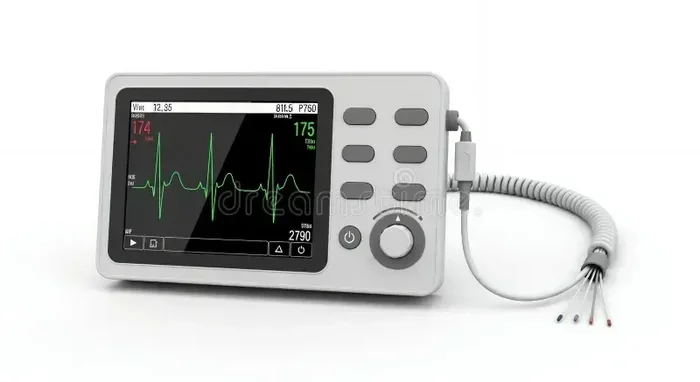
So how does an EKG actually work? The machine itself is not complicated. It has a screen or a paper printout that shows the heart tracing. The electrodes placed on your body send signals to the machine. The machine then amplifies those signals and draws them as waves on a grid.
The grid on an EKG printout has small and large boxes. Each small box represents a tiny amount of time and voltage. This grid helps doctors measure the exact timing of your heartbeat. Without it, reading an EKG would be much harder.
Lead Placement and What It Means
A standard EKG uses 12 different views of your heart. These are called leads. Each lead looks at your heart from a different angle. Proper lead placement is very important. If the electrodes are not in the right spot, the tracing can look wrong. That is why trained technicians carefully place each one.
The 12 leads cover the front, side, and back of your heart. Together, they create a complete picture of your cardiac cycle. Think of it like taking photos from different angles to see a full 3D object. No single lead tells the whole story. But together, they give doctors a clear view.
What Happens During the Test
When you get an EKG, you will lie down on a table. A technician will clean small areas of your skin and place sticky pads on them. These pads are the electrodes. You just need to lie still and breathe normally. The machine does all the work. You should not talk or move during the test. Movement can blur the tracing and make it hard to read.
Some people worry about the EKG meaning being painful. It is not. You will feel a slight stick when the pads go on. After that, you should feel nothing at all. The whole process is over in minutes. Once done, the technician removes the pads, and you are free to go about your day.
Quick Tips for a Smooth EKG Experience
- Wear loose, comfortable clothing that is easy to pull up
- Avoid applying lotions or oils on your chest before the test
- Stay warm, as cold temperatures can tighten your skin and make electrode placement harder
- Tell the technician if you have a pacemaker or sensitive skin
- Remain still and breathe normally throughout the short test
Understanding the Waves and Intervals in an EKG
Now here is where things get interesting. An EKG tracing is made up of distinct waves and intervals. Each part has a specific meaning. Learning what these waves represent is the key to understanding EKG meaning.
The main parts of a normal EKG wave include:
- P wave – This shows the electrical signal that tells the top chambers of your heart to contract. These are called the atria.
- QRS complex – This is the big spike you see on the tracing. It shows the electrical signal that tells the bottom chambers, or ventricles, to contract.
- T wave – This shows the heart resetting itself for the next beat. It represents the recovery phase of the ventricles.
Between these waves, there are important intervals. The PR interval measures the time it takes for the signal to travel from the atria to the ventricles. The QT interval measures the total time it takes for the ventricles to contract and recover. Doctors pay close attention to both of these measurements.
What Each Wave Tells You
Each wave on your heart tracing tells a different part of the story. The P wave is the starting signal. It comes from a group of cells called the sinoatrial node. This is your heart’s natural pacemaker. It sets the pace for every single beat.
The QRS complex is the workhorse of the heartbeat. It shows the ventricles pumping blood out to your body and lungs. If this complex looks too wide or too tall, it could mean something is off. The T wave is the recovery phase. A flat or inverted T wave can be a sign that the heart muscle is not getting enough oxygen.
Understanding these waves helps you see why EKG meaning is so rich. Every bump and dip carries information. A trained eye can spot problems just by looking at the shape and timing of these waves.
Common Mistakes People Make When Reading EKG Traces
Even though you are not expected to read your own EKG, knowing common mistakes helps you understand why a doctor’s interpretation matters. Here are some pitfalls to be aware of:
- Assuming every irregular wave means a serious problem
- Ignoring the importance of lead placement on the final result
- Overlooking how breathing and movement can temporarily change the tracing
- Confusing a single abnormal reading with a chronic condition
- Forgetting that some medications can alter your cardiac rhythm temporarily
What a Normal EKG Tracing Looks Like
A normal EKG tracing follows a predictable pattern. It shows a steady heart rate and regular spacing between beats. The waves have a consistent shape and size. This pattern is called normal sinus rhythm. It means your heart is beating in a healthy, coordinated way.
In a normal tracing, you will see:
- A P wave before every QRS complex
- A steady rhythm with even spacing
- A heart rate between 60 and 100 beats per minute
- A PR interval between 120 and 200 milliseconds
- A QT interval that stays within normal limits
If your tracing looks like this, your heart is likely working well. But remember, a normal EKG does not always mean there are no heart problems. Some conditions may not show up on a resting EKG. That is why doctors sometimes order additional tests.
What Heart Rate Means on an EKG
Your heart rate is one of the first things doctors look at. A resting rate between 60 and 100 beats per minute is considered normal. Athletes often have lower rates, sometimes in the 40s, and that can be perfectly healthy. A rate above 100 at rest is called tachycardia. A rate below 60 is called bradycardia. Both can be normal in certain situations, but they can also signal a problem.
The EKG meaning of your heart rate gives doctors a starting point. If your rate is too fast or too slow, they will dig deeper. They may ask about your symptoms, your medications, or your activity level. Sometimes a simple change in position or breathing can affect your rate too.
Common Abnormal EKG Results and What They Mean
Not every EKG tracing looks perfect. In fact, many people have some kind of abnormality on their EKG. The good news is that not all abnormalities are dangerous. Some are harmless variations. Others may need follow-up testing or treatment.
Arrhythmia and Irregular Heartbeats
An arrhythmia means your heart is beating with an irregular rhythm. This can show up as missing waves, extra waves, or waves that are too far apart. Some arrhythmias are harmless. Others can be serious and need treatment. Common types include atrial fibrillation, premature ventricular contractions, and supraventricular tachycardia.
When an EKG shows an arrhythmia, your doctor will look at your symptoms. Do you feel dizzy? Do you have palpitations? Are you short of breath? The EKG meaning in this case is a clue, not a final answer. Your doctor will combine the tracing with your symptoms and history to make a diagnosis.
Signs of a Heart Attack
One of the most important things an EKG can detect is a heart attack. During a heart attack, blood flow to part of the heart is blocked. This changes the electrical patterns in that area of the heart. The ST segment on the EKG may rise or fall in a way that signals trouble. This is called ST-segment elevation or depression.
If a doctor sees these changes, they will act fast. Quick treatment during a heart attack can save heart muscle and even save lives. This is why EKGs are so commonly used in emergency rooms. The heart attack detection capability of an EKG is one of its greatest strengths.
Other Abnormal Findings
Other things that can show up on an EKG include:
- Enlarged heart chambers, which may show up as taller or wider waves
- Electrolyte imbalances, which can change the shape of the T wave
- Medication effects, since some drugs affect the electrical system of the heart
- Old scar tissue from a past heart injury, which can create unusual wave patterns
These findings do not always mean something is wrong right now. But they do give your doctor useful information. They help guide decisions about your care and monitoring.
Comparison: Normal vs Abnormal EKG Findings
| Feature | Normal EKG | Abnormal EKG |
|---|---|---|
| Heart Rate | 60 to 100 beats per minute | Below 60 or above 100 at rest |
| P Wave | Present before every QRS complex | Missing, inverted, or irregular |
| QRS Complex | Narrow and consistent in shape | Wide, tall, or oddly shaped |
| T Wave | Upright and matching the QRS | Flat, inverted, or peaked |
| ST Segment | Flat and at baseline | Elevated or depressed |
| Rhythm | Regular and steady | Irregular with skipped or extra beats |
| PR Interval | 120 to 200 milliseconds | Too short or too long |
This comparison table gives you a quick visual reference for what doctors look for when they read your tracing. Keep in mind that a single abnormal finding does not always mean a serious problem. Your doctor will look at the full picture before making any conclusions.
When Doctors Order an EKG Test and What to Expect
Doctors order EKGs for many reasons. Sometimes it is a routine check-up. Other times, there are clear symptoms that need investigation. Understanding when an EKG is needed can help you feel more prepared.
Common Reasons for Getting an EKG
Here are some of the most common reasons a doctor might order an EKG:
- You have chest pain or pressure
- You feel dizzy or lightheaded for no clear reason
- You notice your heart racing, fluttering, or skipping beats
- You have shortness of breath during normal activities
- You have a family history of heart disease
- You are starting a new medication that affects the heart
- You are scheduled for surgery and need a pre-op heart check
In each of these cases, the EKG meaning is to give the doctor a quick look at your heart’s electrical health. It helps rule out serious problems or catch them early.
Types of EKG Tests You May Encounter
There are a few different types of EKG tests. A standard resting EKG is the most common. But there are others too:
A Holter monitor is a portable EKG that you wear for 24 to 48 hours. It records your heart tracing continuously as you go about your daily life. This is helpful because a single resting EKG might miss problems that only happen during activity or stress. The Holter monitor gives doctors a longer look at your cardiac rhythm.
There is also a stress EKG, where you walk on a treadmill while the machine records your heart. This shows how your heart handles exercise. If your heart tracing changes during exertion, it could point to blocked arteries or other issues.
Each type of EKG serves a different purpose. Your doctor will choose the right one based on your symptoms and medical history.
EKG vs ECG: What Is the Difference
You may have heard the terms EKG and ECG used interchangeably. They are actually the same test. EKG comes from the German spelling, Elektrokardiogramm. ECG comes from the English version, electrocardiogram. Both terms refer to the same heart monitor procedure. In the United States, EKG is more commonly used. In other parts of the world, ECG is more common. The EKG meaning and ECG meaning are identical. There is no difference in the test itself or the results.
Some people wonder if one is more accurate than the other. The answer is no. Both produce the same electrical activity readings. The only difference is the language origin of the term. So whether your doctor says EKG or ECG, you are getting the exact same test.
Conclusion: Making Sense of Your EKG Tracing
Understanding EKG meaning does not have to be scary or confusing. Your heart tracing is simply a snapshot of the electrical activity that keeps your heart beating. It is a powerful tool that gives doctors valuable information about your cardiac health. Whether your results are normal or show something that needs attention, the EKG is a vital first step.
If you ever get an EKG, remember that those wavy lines tell a story. Each wave, each interval, and each measurement has meaning. A skilled cardiologist can read that story and guide your care. Do not be afraid to ask your doctor questions about your results. Understanding your own heart health puts you in a better position to take care of yourself.
The next time you see an EKG printout, you will know what those lines mean. You will understand the P wave, the QRS complex, and the T wave. You will know what a normal rhythm looks like and why this simple, painless test matters so much. Knowledge is power, and knowing your EKG meaning is one of the best ways to take charge of your heart health.
Frequently Asked Questions
Is an EKG the same as a heart monitor?
Yes and no. An EKG is a specific type of heart recording that captures electrical activity for a short period. A heart monitor is a broader term that can include EKGs, Holter monitors, and event monitors. All EKGs are heart monitoring, but not all heart monitoring is an EKG. The EKG meaning is specifically about recording the heart’s electrical signals for a brief window of time.
Can an EKG detect all heart problems?
No, an EKG cannot catch every heart issue. It is excellent at detecting rhythm problems and signs of a heart attack. But some conditions, like blockages in the arteries, may not show up on a resting EKG. That is why doctors sometimes use stress tests or longer-term monitoring with a Holter monitor to get a fuller picture of your cardiac rhythm.
Is an EKG painful or dangerous?
An EKG is completely painless and very safe. There is no electricity sent into your body. The machine only reads the natural electrical signals your heart already produces. You will feel a slight stick when the adhesive pads are placed and removed, but that is it. There are no risks associated with a standard EKG test.
How long does it take to get EKG results?
A standard EKG takes about 10 minutes to perform. The results can often be read right away by the doctor or technician. In some cases, the tracing is sent to a specialist for a closer look. If your doctor is on-site, you may get your results during the same visit. If not, it could take a day or two for the cardiologist to review and share the findings.
What should I do if my EKG shows something abnormal?
Do not panic. Many abnormal findings are harmless and do not require treatment. Your doctor will look at your results alongside your symptoms and medical history. They may order additional tests like an echocardiogram or blood work to get more information. The EKG meaning is one piece of the puzzle, and your doctor will help you understand the full picture.
How often should I get an EKG done?
There is no set schedule for everyone. Some people get an EKG as part of their annual physical, especially if they have risk factors for heart disease. Others only need one if symptoms arise. Talk to your doctor about what is right for you. If you have a history of heart problems, regular monitoring with an EKG or a heart monitor may be part of your care plan.